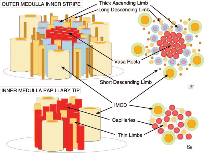
Figure 3. Schematic drawing of outer medulla and papillary tip.

Vasa recta descend in bundles through the inner stripe of the outer medulla (upper panels) surrounded by rings of thick ascending limbs. In the papillary tips, where plaque forms in basement membranes of thin limbs (lower panels), thin limbs are each surrounded by three to four capillaries that derive from the descending vasa recta.
