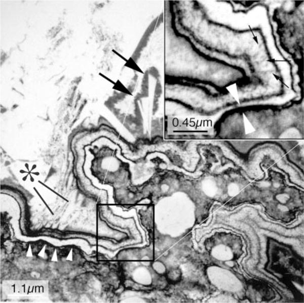
Figure 5. Transmission electron microscopic image of an attachment site of small calcium oxalate (CaOx) stone.

This high-magnification micrograph shows the ultrastructural features of the plaque-stone interface. The upper left hand corner of the micrograph shows the stone material in the urinary space, whereas the lower right corner shows a region of interstitial plaque in the renal papilla. The plaque boundary denoted by a row of four arrowheads has the appearance of a multilayered ribbon. When the portion of this ribbon marked by a square is magnified (see inset at upper right), this ribbon-like structure is seen to have nine separate layers in which five thin black organic lamina alternate with four white lamina. In the thickest of the white lamina, one can see tiny spicules that run perpendicular to the surface and have the appearance of multiple voids that contain tightly packed crystals (small arrows in inset). The two white arrowheads seen in the insert mark the closest black lamina to the renal tissue. An asterisk marks a series of small crystals, whereas two large arrows mark a set of larger crystals that appear to be inserted in the outermost layer (urinary side) of the ribbon. All crystalline material appears to be covered by black matrix material. Adapted from reference 25.
