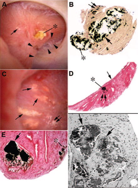
Figure 7. Endoscopic and histologic images of crystalline deposition in brushite stone patients.

Endoscopic examination of papilla from brushite stone-formers reveals a wide range of pathologies from normal to severe deformities. (A) A papilla with extensive pitting marked by a series of arrowheads and extensive dilation of the distal end of ducts of Bellini (BDs) usually filled with a protruding crystalline plug (asterisk). Small regions of Randall’s plaque (single arrow in A) termed “type 1 crystal pattern” were commonly seen. Histologic examination of papillary biopsies reveals enormously dilated inner medullary collecting ducts (IMCDs) and BDs (arrow in B) with mineral occasionally protruding (asterisk in B) in the urinary space. Sites of interstitial plaque are seen (double arrow in B). Other papilla possessed sites of Randall’s plaque (double arrows in C) and regions of yellow plaque (single arrows in C) termed “type 2 crystal pattern.” Each site of yellow plaque was linear in orientation, and when multiple such areas were near each other they formed a spoke- and wheel-like pattern (C). Histologic examination of a single site of yellow plaque revealed these areas represented IMCDs filled with mineral (asterisk in D) that was positioned just beneath the urothelial covering (single arrow in D). Double arrow in panel D marks a site of Randall’s plaque. Light microscopic examination of Yasue-stained sections revealed that the plugged IMCDs contained large amounts of mineral (arrow in E) that filled the lumens of severely damaged tubules whereas nearby tubules appeared normal. Mineral deposits were also seen in thin loops of Henle (asterisk in E). Tissue sections stained with hemotoxylin and eosin revealed crystalline-filled IMCDs (arrows in F) embedded in a region of interstitial fibrosis. Adapted from reference 42.
