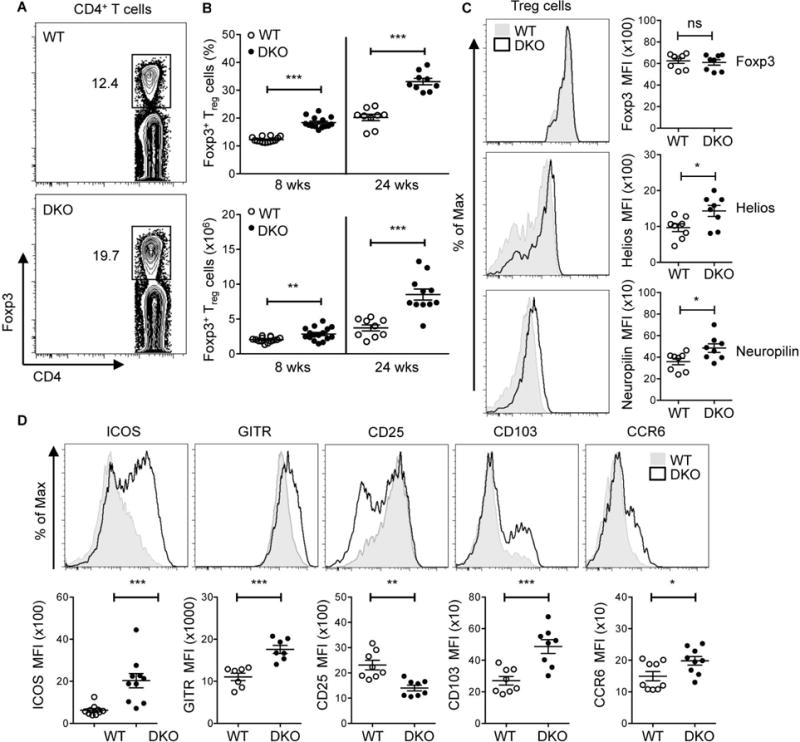
Figure 1. Accumulation of effector Tregs in DKO mice.

A. Representative flow cytometric analysis of the expression of Foxp3 on CD4+ T cells in WT and DKO mice. B. Percentages (top panel) and cell numbers (bottom panel) of CD4+Foxp3+ (Treg) cells in the spleen of 8-12 wks old mice (termed 8 wks) and >24 wks (termed 24 wks) old WT and DKO mice. Data are pooled from at least five independent experiments (8 wks, n=14 and 24 wks, n=9). C. FACS histograms and Mean Fluorescence Intensity (MFI) showing the expression of Foxp3, Helios, and Neuropilin on CD4+Foxp3+ (Treg) cells in the spleen of 8 wks old WT and DKO mice. Representative plots of at least three independent experiments. D. FACS histograms and Mean Fluorescence Intensity (MFI) of Treg cell-associated molecules (ICOS, GITR, CD25, CD103 and CCR6) on CD4+Foxp3+ (Treg) cells from 8-12 wks old WT and DKO spleens. Data are pooled from at least three independent experiments (n=7-10). Each symbol represents an individual mouse. Error bars represent the mean ± s.e.m., *P<0.05, **P<0.01, ***P<0.001 (unpaired, two-tailed Student’s t-test). ns, not significant.
