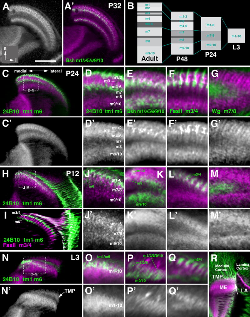
Figure 10.

Layering of the medulla neuropil in the late larva and early pupa. The design of this figure corresponds to that of the preceding Fig.8, presenting frontal sections of medulla neuropil labeled with anti-DNcadherin and layer-specific markers. Top row (A, A′) represents 32h pupa (P32); (B) schematically shows the developmental changes of the medulla neuropil layering as demonstrated with anti-DNcadherin from L3 to adult. Panels of the second and third row (C-G′) represent 24h pupa (P24); rows four and five (H-M′)12h pupa (P12); rows six and seven (N-R) late third instar larva (L3). (R) illustrates the transient medullary plexus (TMP), formed by subsets of medulla neurons, which does not form part of the medulla neuropil. The distal boundary of this neuropil is defined by the incoming lamina/retina axons, visible as a DNcadherinpoor band (arrow). For detail, see text. Bar: 50 μm (A, C, H, I, N)
