
Figure 11.
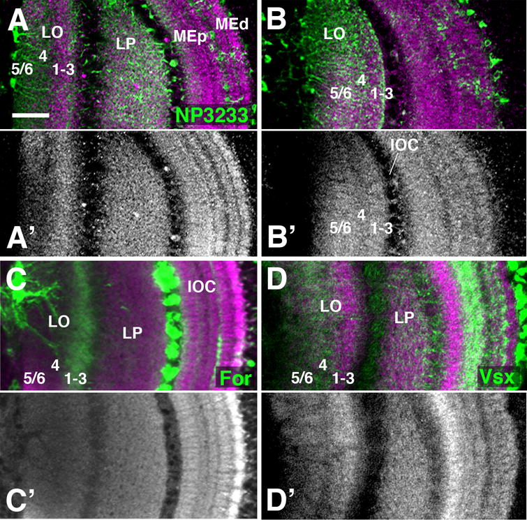
Layering of the late pupal lobula neuropil. (A-D′). Frontal sections of P72 optic lobe labeled with anti-DNcadherin (magenta in A-D; gray in A′-D′), and co-labeled with NP3233-Gal4 [expressed in glia; green in (A,B)]; for-Gal4 [lineage tracing; labels T neurons; green in (C)]; and Vsx1-Gal4 [labels Tm neurons; green in (D)]. (B, B′) represent a plane of section anterior to that in (A, A′), showing only the lobula (LO) and not the lobula plate (LP). The DNcadherin expression in the lobula neuropil reveals three layers, including a central layer with moderate signal intensity, flanked by a proximal and distal layer with higher intensity. Processes of neuropil glia occur at a slightly higher density in the intermediate layer than the flanking proximal and distal layers (B). The distal layer contains terminals of T neurons (C), identifying this wide band as combined lobula layer 1-3 (Fischbach and Dittrich, 1989). Vsx1-positive Tm neurons are confined to the proximal and intermediate layers, indicating that they comprise lobula layers 4-6, which receive the large majority of Tm/Tmy neurons (Dittrich and Fischbach, 1989). The lobula plate (LP) is uniformly labeled by DNcadherin and there appears to be no clear layer separation with the present markers. For other abbreviations, see legend of Fig.2. Bar: 25 μm