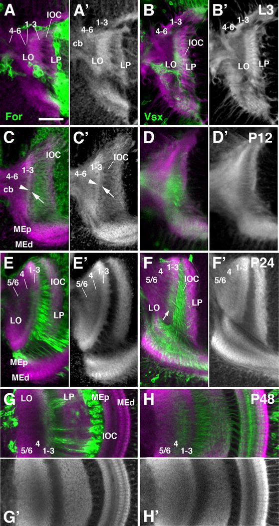
Figure 12.

Layering of the lobula neuropil during metamorphosis. Frontal sections of the optic lobe labeled by for-Gal4 lineage tracing (green; A-A′, C-C′, E-E′, G-G′) and Vsx1-Gal4 (green; B-B′, D-D′, F-F′, H-H′) and counterstained with anti-DNcadherin (magenta in A-H; gray in A′-H′) at late larval stage (L3; first row; A-B′), 12h pupa (P12; second row; C-D′), 24h pupa (P24; third row; E-F′), and 48h pupa (P48; fourth and fifth row; G-H′). The tripartite subdivision of the lobula neuropil is visible from P24 onward (E, E′). Prior to this stage, a thin, DNcadherinrich band demarcates a protolayer for 1-3, in which for-Gal4-positive axon terminals are concentrated [arrowhead in (C)]; further proximally, the nascent lobula neuropil, which is in broad contact with the central brain neuropil (cb), has a moderate DNcadherin signal [protolayer 4-6 in (A-C′)]. Note that Vsx1-positive Tm axons, concentrated in proximal layers 4-6 in the adult lobula (see Fig.10D), are in the intermediate layer 4 at P48, and in distal layers 1-3 prior to that [arrow in (F)]. White arrows in (C, C′) point at superficial band of moderate DNcadherin signal that is continuous with the striated pattern of fiber bundles forming the inner optic chiasm (IOC). This band could correspond to layer of glial cells associated with the IOC. For abbreviations, see legend of figure 2. Bar: 25 μm
