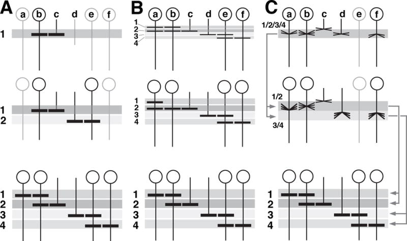
Figure 13.

Hypothetical mechanisms of neuropil layer formation. (A-C) show time series (top: early stage; bottom: late stage) of schematic cross sections of layered neuropil, such as the medulla. Shaded horizontal bars (1-4) represent layers. Vertical elements (a-f) represent interneurons (a, b, e, f) and photoreceptor afferents (c, d) whose terminal branches (horizontal lines within layers) generate the layers. (A) Layers are formed sequentially. Elements b and c generate the top layer (1). Only these elements differentiate early (top). As a result, the only layer present at early stage is (1). Other elements and their layers follow at subsequent stages. (B) Layers form simultaneously; development merely consists in increase in branching/layer thickness. (C) Layers develop from protolayers. Most interneurons and afferents appear around the same stage (top) and form undifferentiated terminal branches that largely overlap in a protolayer (1/2/3/4). Protolayers may already be polarized, with elements later restricted to deep layers [e.g., (f)] occupying a deeper position within protolayer, and vice versa. Some elements (c, e) may not initially form part of protolayers.
