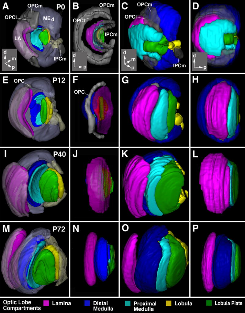
Figure 5.

Morphogenesis of the optic lobe. All panels show digital 3D models at the pupariation (P0; first row, A-D), 12h after puparium formation (P12; second row, E-H), 40h after puparium formation (P40; thrird row, I-L), and 72h after puparium formation (P72; bottom row, M-P). Panels of first column (A, E, I, M) and third column (B, F, J, N) present view from dorso-lateral posterior; second column (B, F, J, N) and fourth column (D, H, L, P) show lateral view. Insets at lower right corner of panels of first row (A-D) delineate orientation of panels (a anterior; d dorsal; l lateral; p posterior). In panels of first and second column, developing neuropils are shown in saturated colors, using the color code indicated at bottom of figure. In these two columns, the epithelial part of the outer and inner proliferation centers (OPC, IPC) is in gray. In panels of first column, cortices of the lobula plate/proximal medulla are omitted to permit view of the neuropils. The cortex of the distal medulla (MEd) and lamina (LA) is rendered in semi-transparent, unsaturated colors. In second column, all cortices are omitted. In third and fourth column, cortices are shown in saturated colours.
