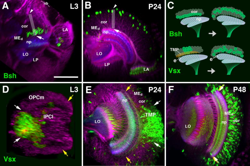
Figure 8.

Cell body movements in the medulla cortex. (A, B) Horizontal confocal sections of optic lobe of late larva (L3, A) and early pupa (P24, B). Medulla intrinsic neurons (Mi1) are labeled by bsh-Gal4 (green); general labeling of neurons by anti-Neuroglian (red) and neuropil by anti-DNcadherin (blue). The vertical axis of the medulla cortex is shown by gray rectangle. Shortly after their birth, Mi1 cell bodies are close to medulla neuropil [arrow in (A)]; after 24h, they have shifted to distally towards the outer surface of the medulla cortex [arrowhead in (B)]. Top part of panel of (C), showing a schematic frontal section of medulla cortex and neuropil, schematically depict the cell body movement. (D-F) Confocal sections of optic lobe of late third instar (L3, D), early pupa (P24, E) and mid-stage pupa (P48, F). Labeling of Tm neurons by Vsx1-Gal4 (green). General labeling of neurons by anti-Neuroglian (red) and neuropil by anti-DNcadherin (blue). Vsx1-positive neurons originate from a central domain of the outer proliferation center [OPCm; in between white arrows in (D)], whereas the dorsal and ventral wings of the OPCm (yellow arrows) are devoid of this cell type. The same spatial restriction is still visible at P24 (E), where Vsx1-positive cell bodies are located in the central part of the cortex. Note that axons of the neurons at the dorsal rim of the Vsx1-positive cluster (white arrow) project dorsally, traverse the cortex and enter the neuropil at its very dorsal edge (yellow arrow); likewise, neurons at the ventral rim project ventrally. The mass of these fibers, which fill the deeper part of the medulla cortex, form the transient medullary fiber plexus (TMP; see Fig.6B and 9R). By P48 (F), cell bodies have spread out to fill the entire cortex (white arrows and yellow arrows coincide). All Tm axons extend parallel, and the TMP has disappeared. The movement of cell bodies is depicted schematically in bottom part of panel (C). The entry point of a representative Vsx1-positive axon, indicated by “e”, remains the same before and after the movement of cell bodies. For other abbreviations see legend of Fig.2. Bar: 50 μm
