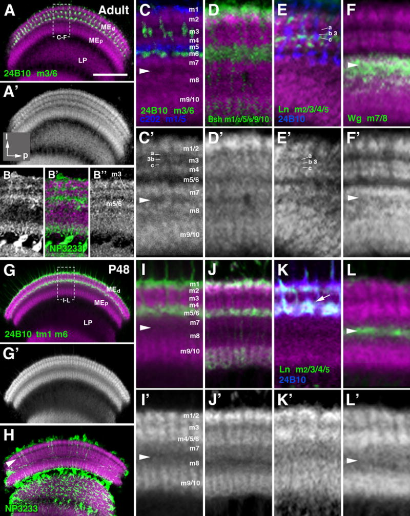
Figure 9.

Layering of the medulla neuropil in the adult and mid-stage pupa. (A-L′) Frontal sections of medulla neuropile, labeled by anti-DNcadherin (magenta in A-L; white in A′, B″, C′-L′), which reveals distinct strata with different signal intensity. Double labeling with cell type-specific markers, traditionally used to define layers m1-m10, permits one to assign the DNcadherin pattern to these layers. The layer-specific markers used are: anti-Chaoptin (24B10; labeling of photoreceptors R7/8; green in A, C, I; blue in E, K); c202-Gal4 (labeling of lamina L1 neurons; blue in C); bsh-Gal4 (labels medulla Mi6 neurons; green in D, J); ln-Gal4 (labels lamina L3/4 neurons; green in E, K); wg-Gal4 (labels precursors of medulla tangential neurons; green in F, L). The medulla layers (m1-10) defined by the specifically neuronal populations are indicated next to the marker (lower left of panels (A-F) and (G, K). Note that anti-Chaoptin labels adult layers m3 and m6; at P8 and earlier (see Fig.9) it labels m6 and transient layer distal of m1 (“tm1”). The top two rows of panels (A-F′) show adult stage; bottom rows (G-L′) pupa at 48h after puparium formation (P48). For a given pair of panels (e.g., C, C′), the upper one shows anti-DNcadherin label (magenta) in conjunction with marker; the lower one is anti-DNcadherin only (in white). Panels of left column (A, A′, G, G′) present overview of medulla neuropil at low magnification; hatched lines outline domain of neuropil shown at high magnification in the four columns to the right. Panels (B-B″) and (H) show labeling of neuropil glia expressing the marker NP3233-Gal4 (green). White arrowhead in (C, C′, F, F′, I, I′, L, L′) indicates serpentine layer (boundary region between m7 and m8. White arrowhead in (H) marks the DNcadherinlow stratum (nascent layer m3) prior to the formation of significant glial processes. Note differences in anti-DNcadherin pattern between late pupal and adult stage illustrated in panels (C′) and (I′), respectively. Layer m3, in the adult, is represented by a thin band with moderate DNcadherin signal (“3b”) flanked by bands of low signal (“m3a”, “m3c”). In the 48h pupa, m3 is narrower and typically represented by a single band with low or moderate DNcadherin level. Most conspicuously, the DNcadherinlow band representing layers m5/6 in the adult (C, C′) is absent in the pupa (I, I′); here, terminals of m5/6 specific neurons (L1, R8), that coincide with a DNcadherinlow stratum in the adult, are found in the lower part of a DNcadherinhigh band (I). This band represents the “protolayer” for m4-m6. For other abbreviations, see legend of Fig.2. Bar: 50 μm (A, G)
