
Fig. 1. Refined neutron diffraction data taken at 5 K.
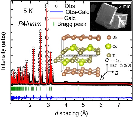
Small impurity peaks were excluded from the refinement. The upper inset shows a scanning electron microscopy image of a typical crystal of CeSbTe, and the lower inset shows a drawing of the crystal structure of CeSbTe, where the nonsymmorphic symmetry elements are highlighted. arbs, arbitrary units; Obs, observed; Calc, calculated.