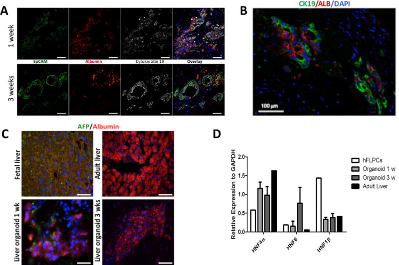
Figure 1. Lineage specification of human fetal liver progenitor cells (hFLPCs) and formation of liver organoids.

A) Distribution and phenotypic characteristics of hFLPCs during 1 (top panel) and 3 (bottom panel) weeks of differentiation in culture. Cells were stained for epithelial cell adhesion molecule (EpCAM), albumin (ALB), cytokeratin19 (CK19) and for cell nuclei (DAPI). Scale bar is 20 μm. B) Immunostaining of liver organoids after 3 weeks of differentiation shows ductal structures containing cells expressing both CK19 (green) and albumin (red). C) Expression of α-fetoprotein (AFP, green) and albumin (ALB, red) in liver organoids after 1 and 3 weeks differentiation and in fetal and adult liver tissues. Scale bar is 20 μm. D) RT-PCR analysis of the expression of hepatic transcription factors hepatocyte nuclear factor (HNF) 4α, HNF6 and HNF1β in freshly isolated hFLPCs, liver organoids after 1 and 3 weeks differentiation, and in adult liver tissue.
