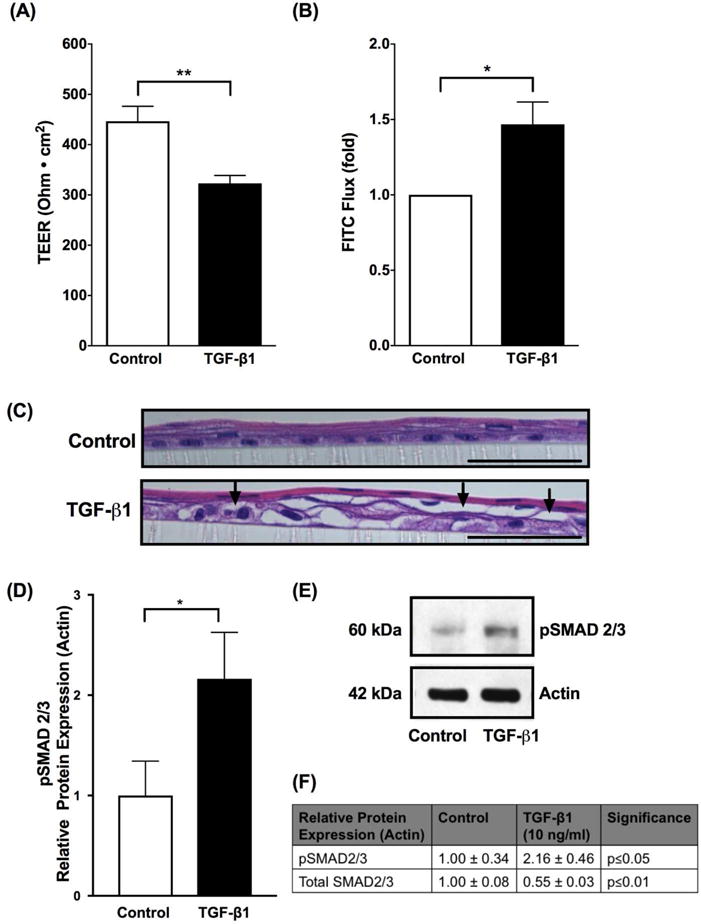
Figure 1. TGF-β1 diminishes esophageal epithelial barrier.

Using immortalized human esophageal epithelial (EPC2-TERT) cells in a stratified squamous 3-dimensional air liquid interface (3D-ALI) culture, transforming growth factor-β1 (TGF-β1) (10 ng/ml) diminished barrier as measured by (A) transepithelial electrical resistance (TEER) and increased permeability as measured by (B) 3kDa FITC dextran paracellular flux (FITC flux) (N=4–8, *p≤0.05, **p≤0.01). (C) H&E stained sections from EPC2-TERT cells grown in 3D-ALI culture and treated with TGF-β1 (10 ng/ml). Arrows indicate prominent cellular separation. Black scale bar represents 50 μm. (D) To confirm that TGF-β1 signaling is increased, we performed western blot densitometry which shows increase in protein expression of phosphorylated SMAD2/3 (pSMAD2/3) when cells are exposed to TGF-β1 (10 ng/ml), indicating that TGF-β1 activates SMAD2/3 via phosphorylation in the in vitro 3-D ALI model (N=4, *p≤0.05). (E) Representative western blot showing increase in protein expression of pSMAD2/3 in vitro when exposed to TGF-β1 (10 ng/ml). (F) Protein expression of pSMAD2/3 and total SMAD2/3 when EPC2-TERT cells exposed to TGF-β1 (10 ng/ml). Data are expressed as mean fold change versus unstimulated controls ± SEM. Treatment with recombinant human TGF-β1 (10 ng/ml) occurred at the start of 3D-ALI exposure and during the process of differentiation and stratification on days 7 and 9.
