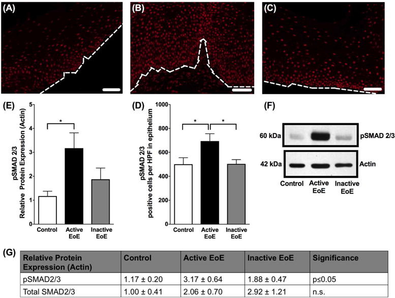
Figure 7. pSMAD2/3 in pediatric subjects.

Immunofluorescent staining of phosphorylated SMAD2/3 (pSMAD2/3) in human esophageal biopsies from (A) control (B) active eosinophilic esophagitis (EoE) and (C) inactive EoE subjects. A C at 200× magnification, white scale bar represents 50 μm. White dotted line represents basal lamina. (D) Quantification shows increased positive cells per high power field of pSMAD2/3 staining in active EoE compared to control and inactive EoE. (E) Western blot densitometry shows increase in protein expression of pSMAD2/3 in active EoE vs control and inactive EoE. (F) Western blot showing increase in protein expression of pSMAD2/3 in active EoE vs control and inactive EoE. (G) Relative protein expression of pSMAD2/3 and total SMAD2/3 in active EoE vs control and inactive EoE.
