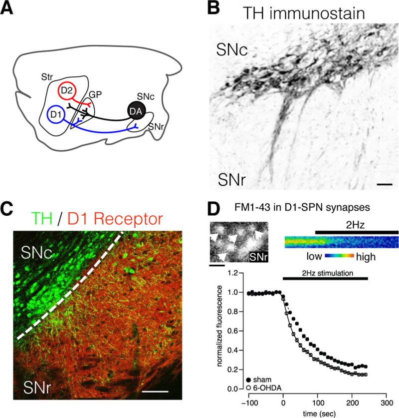
Figure 1. Modulation of SPN neuronal plasticity occurs at multiple synaptic sites.

A) Simplified schematic of striatofugal SPN and ascending DA projections in the rodent brain. DA neurons of the SNc can influence neuronal plasticity in distinct microcircuits via post- and presynaptic DA receptors in the striatum, GP and SNr. B) Immunolabel for tyrosine hydroxylase (TH) in mouse ventral midbrain showing DA neurons of the SNc and their dendritic processes extending into the SNr. Somatodendritically released DA presumably influences motor functions by local actions within the midbrain. Scale = 50 μm. C) Immunolabel for TH (green) and D1R (red) in a mouse midbrain section. D1R are preferentially located on the terminals of D1-SPNs, and thus DA neurons directly influence local GABA release from the striatonigral direct pathway. Scale = 100 μm. D) Labeling of D1-SPNs synapses with the fluorescent vesicular probe FM1–43 to study presynaptic plasticity produced by DA denervation. Upper panels show 2-photon images of FM1-43-labeled D1-SPN synaptic boutons in the SNr (left), and a time-series of destaining from a single fluorescent bouton during stimulation of D1-SPN projections (right). Loss of fluorescence represents the fusion of GABA-containing vesicles during electrically evoked synaptic activity and the rate of decay reveals the probability of neurotransmitter release. Bottom panel shows the normalized FM1-43 fluorescence as a function of time from >100 D1-SPN synaptic boutons in brain slice preparations from DA intact (sham) and denervated parkinsonian mice (6-OHDA). The rate of vesicular fusion is greatly increased in DA denervated animals, indicating that increased GABA release during synaptic activity provides a potential mechanism for LID. Scale = 3 μm.
