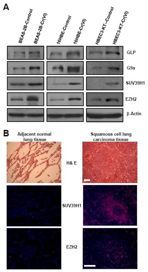
Fig. 2. The expression levels of several histone-lysine N-methyltransferases (HMTases) are increased in Cr(VI)-transformed cells and Cr(VI) exposure-caused human lung cancer tissues.
(A) Representative Western blot images for the analysis of several HMTases levels in passage-matched control and Cr(VI)-exposed human bronchial epithelial. After exposure to 0.25 μM of Cr(VI) (K2Cr2O7) for 20 (BEAS-2B), 40 (16HBE) and 30 (HBEC3-KT) weeks, passage- matched control and Cr(VI)-exposed BEAS-2B, 16 HBE and HBEC3-KT cells were harvested for Western blot analysis as described in Methods. Similar results were obtained in two additional experiments. (B) Representative images of H&E staining and immunofluorescence (IF) staining of SUV39H1 and EZH2 in the squamous cell lung carcinoma tissue and the adjacent normal lung tissue from a non-smoker worker exposed to chromate for 19 years. Similar staining results were obtained in lung cancer tissue from another non-smoker worker exposed chromate for 38 years. Scale bar: 100 μm.

