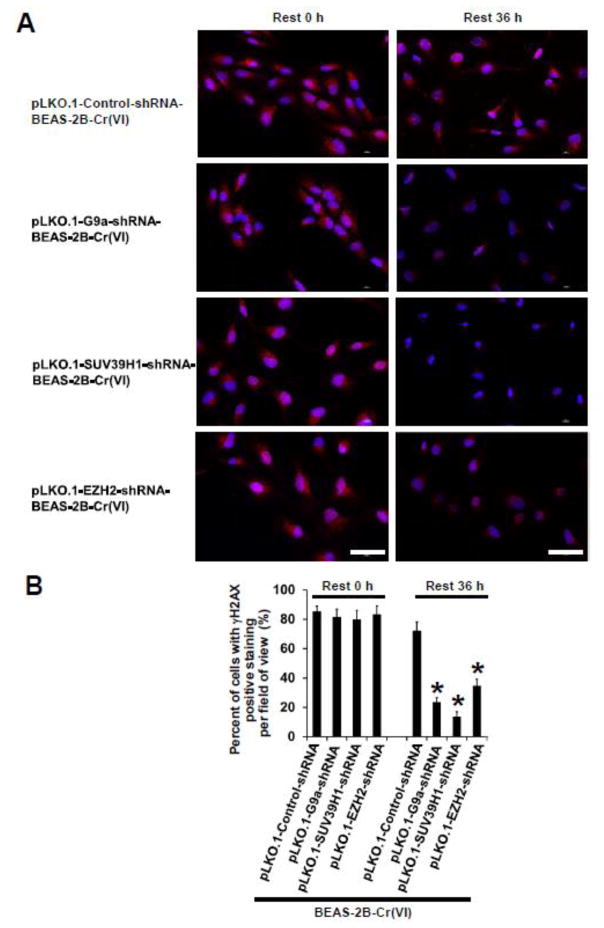
Fig. 5. Stable knockdown of HMTases significantly reduces Cr(VI) exposure -caused DNA damage.
(A) Representative images of immunofluorescence (IF) staining of γH2AX in chronic low dose Cr(VI)-exposed shRNA vector control and each HMTase shRNA stable knockdown BEAS-2B cells treated with 2.5 μM of Cr(VI) (K2Cr2O7). After exposure to 0.125 μM of Cr(VI) (K2Cr2O7) for 25 weeks, vector control and each HMTase stable knockdown cells were seeded in 6-well plates. After overnight culture, cells were treated with 2.5 μM of K 2Cr2O7 for 12 h. At the end of the treatment, one set of cells were used for IF staining of γH2AX (designated as Rest 0 h). The other set of cells were washed with PBS and cultured for additional 36 h in the absence of K2Cr2O7 and used for IF staining of γH2AX (designated as Rest 36 h) as described in Methods. Scale bar: 100 μm. (B) Quantitation of γH2AX positive staining in Cr(VI)-exposed cells described in (A). Results are expressed as percent (%) of γH2AX positive staining per field of view. Data are presented as mean ± SD (n=30 fields of view). *p< 0.05, compared to Cr(VI)-exposed shRNA vector control cells. Similar results were obtained in two additional experiments.

