
Figure 4.
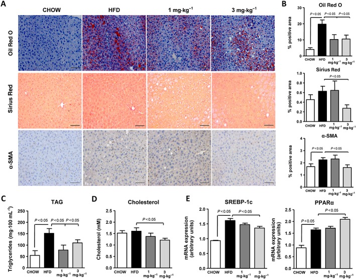
Effects of IW‐1973 on hepatic steatosis and fibrosis in the HFD obesity model. (A) Representative photomicrographs (200× magnification) of liver sections stained with Oil Red O, Sirius Red and α‐SMA antibody in mice receiving chow diet (n = 5), an obesogenic HFD diet (n = 10), HFD plus sGC stimulator IW‐1973 at 1 mg·kg−1 (n = 10) or HFD plus IW‐1973 at 3 mg·kg−1 (n = 10). (B) Histomorphometric analysis of the area stained with Oil Red O, Sirius Red and α‐SMA. (C) Hepatic TAG levels. (D) Hepatic cholesterol levels. (E) Hepatic SREBP‐1c and PPARα mRNA expression. Results are expressed as mean ± SEM. Scale bar = 50 μm.