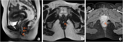
Fig. 3.

Anovaginal fistula. A thick gauge fistula exibited abscess formation involving the recto vaginal septum (arrows). a Sagittal T2-weighted image. b Oblique-transverse T2-weighted image. c Post-gadolinium oblique-transverse fat-supressed gradient echo T1-weighted image
