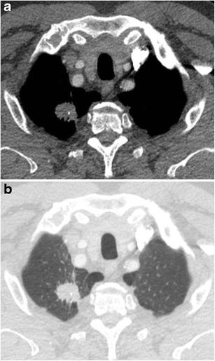
Fig. 17.

Axial CT-images in lung (a) and mediastinal (b) window setting show a 2.4 cm nodule in the apex of the right upper lobe. The lesion is somewhat spiculated with scarse small dot-like calcifications. Although postinfectious scarring is common in the lung apices, there are no abnormalities on the left. Moreover the lesion is relatively round and the calcifications do not have a typical benign nature. Histopathologic examination after lobectomy showed a moderately differentiated squamous cell carcinoma
