
Fig. 8.
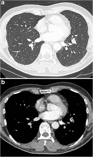
Incidental finding of a solitary pulmonary nodule in a 50-year-old man. Axial CT-image in lung window setting a shows a well-delineated nodule with clear lobulated morphology in the left upper lobe. The absence of growth, internal benign-looking calcifications and clear hypodense areas (corresponding to fat) on the images in mediastinal window setting b led to the probable diagnosis of a hamartoma