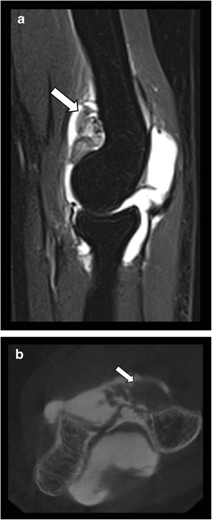
Fig. 12.

Pigmented villonodular synovitis of the elbow. a. Sagittal T2–WI fat suppressed MRI image presenting a mass within the joint cavity (arrow) with a low signal areas related to hemosiderin deposits. b. CBCT-A showing the presence of proliferative synovium (arrow) within the anterior part of the joint cavity
