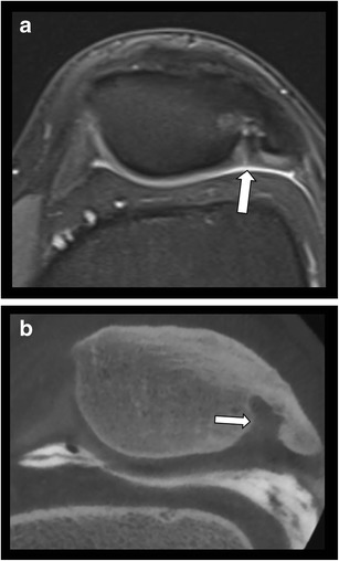
Fig. 13.

Dorsal defect of the patella simulating a large cartilage defect on MRI. a. Axial T2 WI fat saturated MR image shows a focal bony defect with surrounding bone marrow edema at the superolateral aspect of the patella. There is suspicion of an overlying cartilage fissure (arrow). b. Axial reformatted CBCT-A demonstrates a dorsal patella defect. The overlying patellar cartilage is intact.
