
Fig. 2.
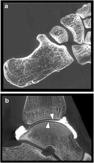
Evaluation of bone architecture and normal articular cartilage. a. Axial CBCT image of a cadaver foot illustrating exquisite detail of the cortical and trabecular bone architecture. b. Sagittal reformatted image of a CBCT-A of the talocrural joint showing smooth surface of normal articular cartilage surface of distal tibia and talar dome (arrowheads)