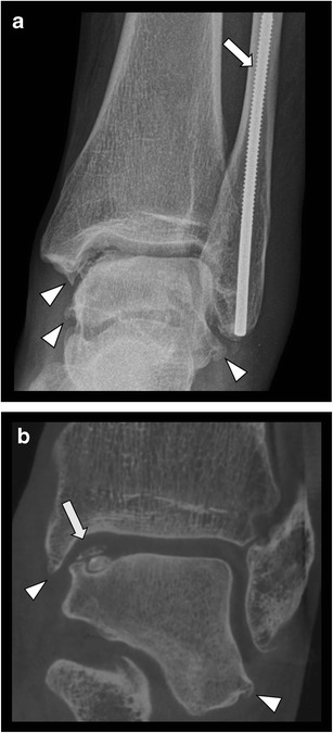
Fig. 9.

Posttraumatic unstable osteochondral lesion of the talus and massive degenerative changes of the ankle in a 66-year-old female. a. CR (AP view) demonstrating the presence of metallic screw within the fibula (arrow). Note irregular articular surface of talar dome and advanced degenerative changes with osteophytes formation (arrowheads). b. CBCT coronal reformatted image better shows the presence and extent of an unstable osteochondral lesion (arrow) and osteophytes (arrowheads). There is no metal artifact from the screw in the fibula
