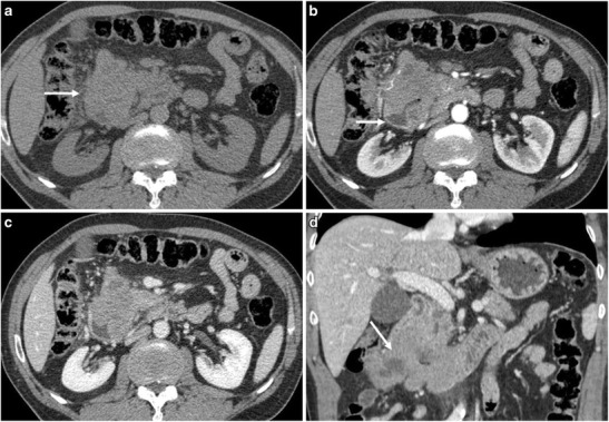
Fig. 2.

A 62-year-old male patient with primary pancreatic lymphoma, histotype high-grade B-cell lymphoma not otherwise specified (Ki 67 score > 90%). a CT scan of the abdomen shows the presence of a solid mass in the pancreatic head (arrow). b, c, d CT images after contrast medium administration in the arterial-pancreatic phase (a) and portal-venous phase (b, c): the neoplasm protrudes in the duodenum (arrow), as better demonstrated on coronal reconstruction (d). The histopathological diagnosis was reached with endoscopic biopsy of the involved duodenal wall
