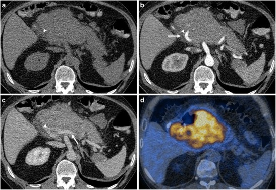
Fig. 3.

A 51-year-old male patient with primary pancreatic lymphoma, histotype diffuse large B-cell (Ki 67 score > 90%). a CT scan of the abdomen shows the presence of a large solid mass in the body-head of the pancreas. A biliary stent is seen. b, c CT images after contrast medium administration in the arterial-pancreatic phase (a) and portal-venous phase (b): the arteries and veins (arrows) are encased but not infiltrated. The main pancreatic duct is not enlarged. d This 18FDG PET-CT fusion image reveals high metabolic activity in the lesion
