
Fig. 4.
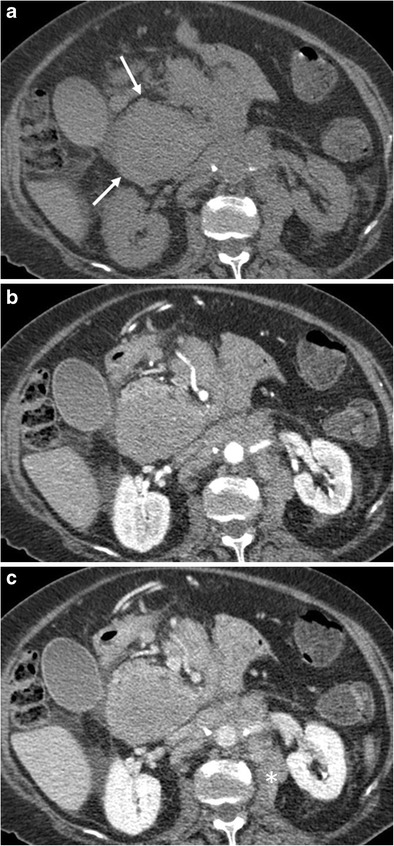
A 79-year-old female patient with primary pancreatic follicular lymphoma (Ki 67 score < 25%). a Unenhanced CT scan depicts a hypodense mass in the pancreatic head (arrows). b, c CT images after contrast medium administration in the arterial-pancreatic (b) and portal-venous phase (c): progressive enhancement of the neoplasm is visible. Enlarged retroperitoneal lymph nodes with ill-defined margins are depicted (*)