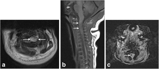
Fig. 12. Spinal cord injury in 4-day-old female with a history of shoulder dystocia presenting with right sided upper and lower extremity neurologic deficits.

Axial T2 (a) and sagittal T1 (b) images of the cervical spine demonstrate a focal area of T1 hyperintensity and T2 hypointensity (arrows) in the high cervical cord at the level of C2–3 consistent with acute injury. Focal hemorrhage in the right cervical cord with susceptibility artifact seen on C (axial SWI). Finding was thought consistent with spinal cord injury secondary to stretching/traction
