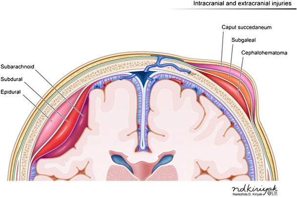
Fig. 2.

Illustration depicting hemorrhages by location within the different layers of the meninges (left of image) and scalp (right of image)

Illustration depicting hemorrhages by location within the different layers of the meninges (left of image) and scalp (right of image)