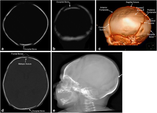
Fig. 5. Molding of the skull post vaginal delivery.

Immediate post-delivery appearances of the skull on head CT. The occipital bone is slightly depressed with associated sutural overlap as seen on the axial and coronal CT images and the 3-D reconstructions (a-d). Lateral skull radiograph (e) demonstrates overlap of the occipital bone (white arrow)
