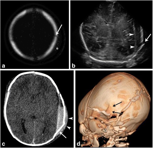
Fig. 6. Skull fractures in two neonates.

Axial bony algorithm reconstruction from a head CT (a) in a 2-day-old demonstrates a non-displaced linear left parietal skull fracture (arrow) with overlying soft tissue swelling (marked by asterisk on a). Ultrasound and CT images on another 1-day-old male (b, c) with a history of traumatic delivery characterized by multiple attempts of vacuum extraction. Coronal gray scale ultrasound image (b) demonstrates a displaced left parietal fracture (arrow) with underlying extra-axial fluid collection (arrowheads). Axial non-contrast head CT image (c) shows a complex left parietal bone fracture with an angulated anterior component and an adjacent depressed “ping-pong” fracture component posteriorly (arrow). There is an associated overlying hyperdense fluid collection consistent with cephalhematoma (arrowhead). There is also an underlying large epidural hemorrhage with fluid/fluid levels. 3-D volume rendered image (d) re-demonstrates the complex left parietal bone fracture (black arrows)
