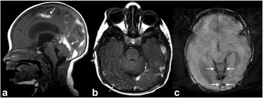
Fig. 8. Subdural Hematoma.

Brain MRI in a 6-day-old term neonate with history of vacuum-assisted delivery. Sagittal T1 (a) MR image demonstrates subdural hematomas tracking along bilateral occipital lobes and along the tentorium. Another 8-day-old term neonate with history of prolonged rupture of membranes shows subdural hemorrhage (b) layering along the occipital-parietal convexities and along the tentorium (arrows). Corresponding axial gradient-recalled echo (susceptibility-weighted) image (c) at a slightly more cephalad level reveals hemosiderin staining along the tentorium and posterior convexities of the brain (arrows)
