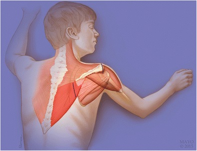
Fig. 4.

Origin and insertion of the trapezius muscle, highlighting the lower trapezius. Incisions for the open lower trapezius transfer are shown with solid lines. A 5-cm incision is made 1-cm medial to the medial scapular spine as shown here. The tendon will be passed through the area designated by the dashed line. Note the close proximity of the spinal accessory nerve (CN XI) to the incision. The nerve lies below the fascia of the trapezius, making superficial dissection in this region safe. Used with permission of Mayo Foundation for Medical Education and Research. All rights reserved
