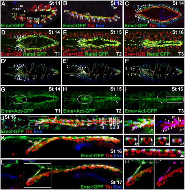
Fig. 1.
Diversity of EPCs and formation of the OHS. (A,B) Lateral views from Eme>mcd8GFP stage 11 (A) and 12 (B) embryos showing 12 EPC clusters in dorsal mesoderm. The parasegments (PSs) from which OHS EPCs originate are coloured yellow. Asterisk in A indicates missing EPCs in PS13. (C) Dorsal view of a stage 14 embryo. Arrowheads indicate the OHS EPCs from PS2 and PS3; asterisks indicate detaching WH EPCs. The OHS EPCs from PS6 are indicated by arrows. (D-F′) Dorsal views of stage 14-16 living Hand-nlsGFP;Eme>H2B embryos showing cell tracking (lines) of migrating EPCs (white or yellow dots). (G-I) Dorsal views of Eme>lifeAct-GFP stage 14-16 living embryos. Leading edge activity of the migrating OHS EPCs is increased (G, circled). As the heart closure proceeds (H) the OHS EPCs become aligned (circled) with the apparent actin cable (arrow), whereas the heart proper EPCs (arrowheads) form a network of interconnected cells. At stage 16 (I), the actin cables (arrows) link the aorta EPCs, whereas the heart proper-associated EPCs (rectangle) display a patched actin enrichment. (J) Dorsal view of a stage 16 Eme>mcd8GFP embryo. (J1,J1ʹ) The OHS EPCs appear highly compacted and located dorsally above the heart tip. The EPCs associated with the aorta, except the first pair (arrowhead), lie more laterally and are interspaced according to the segmental register. The heart proper EPCs are located above or lateral to the cardioblasts (J2-J6), and appear as a dorsally connected network of cells. (K,L) Lateral views of stage 16 (K) and 17 (L) embryos stained as above and showing the arrangement of the heart-associated EPCs, which are interconnected. The OHS EPCs (L1,L1′) make a link between the bent anterior aorta and the epidermis.

