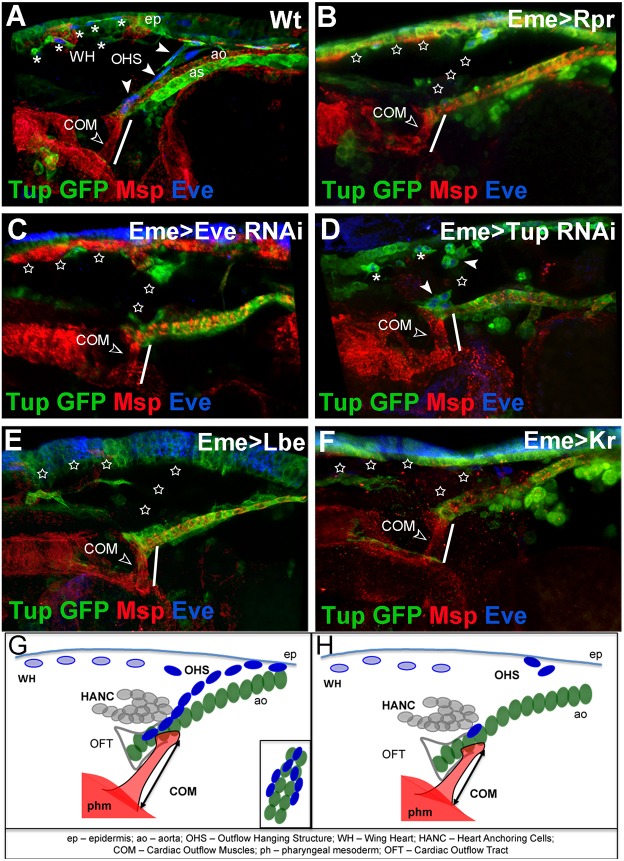
Fig. 3.
The OHS is required for cardiac outflow positioning. (A-F) Lateral views of the cardiac outflow region from Tup-GFP late stage 16 embryos. Tup-GFP reveals EPCs, aorta (ao), amnioserosa (as) and epidermis (ep); Msp300 stains COMs (open arrowhead) and cardiac cells; and Eve labels EPCs. (A) Wild-type embryo: the OHS EPCs are indicated by arrowheads and WH EPCs by asterisks. (B) Eme>Rpr embryo in which EPCs were ablated (stars). The COMs appear shorter (white line) than in wild type (compare A with B). (C) EPC-specific eveRNAi results in partial loss of OHS and WH EPCs (stars), and affects COMs length (white line). (D) EPC-targeted tupRNAi results in partial loss of EPCs. The COMs appear shorter (white line). (E,F) The eme-driven misexpression of Lbe (E) or Kr (F) represses eve, leading to affected specification of OHS and WH EPCs, and shortening of COMs (white lines). (G) Cellular components of cardiac outflow, including OHS. The OHS EPCs (dark blue) connect the tip of the anterior aorta (green) to the epidermis and, like COMs, are attached on both sides of the cardiac outflow. (H) Affected positioning of the cardiac outflow associated with COM shortening in the contexts where the OHS is lost.

