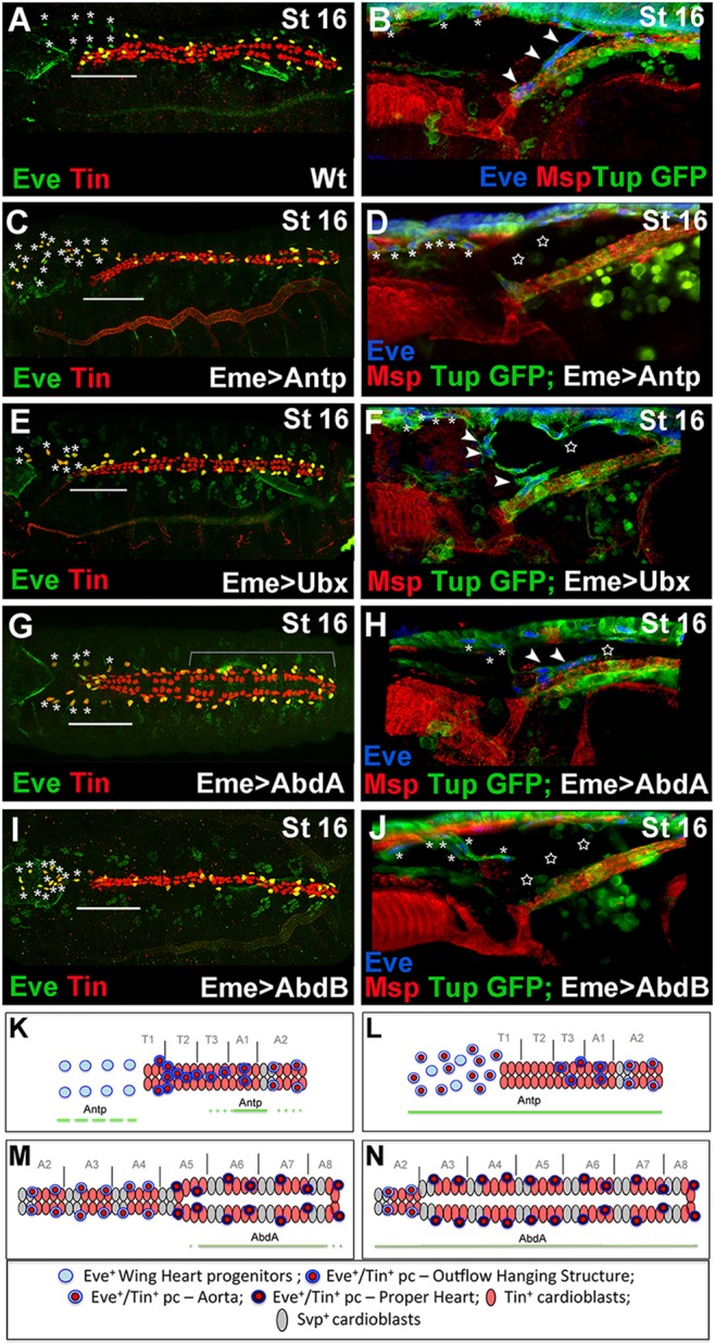
Fig. 4.

Hox genes control EPCs diversity. (A,C,E,G,I) Dorsal views of late stage 16 embryos stained with Tin and Eve. (B,D,F,H,J) Lateral views of the cardiac outflow region from late stage 16 Tup-GFP embryos stained for GFP, Eve and Msp300. (A,B) In the wild-type embryos, the OHS EPCs are associated with the anterior aorta (A, above the line; B, arrowheads) and the WH EPCs are located anteriorly to the heart (asterisks). (C,D) In the Eme>Antp context, an increased number of EPCs adopt the WH fate (C,D, asterisks) and the OHS EPCs are almost absent (C, above the line; D, stars). (E,F) Eme-targeted overexpression of Ubx disrupts OHS morphogenesis (F, star) without shifting the identity of OHS EPCs to WH fate (E, above the line). Few OHS EPCs (arrowheads) are located more anteriorly compared with wild type (A). Asterisks indicate WH cells. The heart proper appears thinner. (G,H) In eme>AbdA embryos, the WH EPCs (asterisks) migrate more slowly, the string of OHS EPCs is broken (F, star) and the aorta is enlarged (G, bracket). (I,J) The Eme-driven expression of AbdB leads to a reduced EPC number. The OHS is missing (J, stars) and a few persisting EPCs adopt the WH fate (I, asterisks). In all EPC-targeted Hox misexpression experiments, the specification of the OHS EPCs is affected and the WH EPCs do not lose Tin expression. (K) Anterior aorta and WH progenitors in wild-type context. Solid and broken lines indicate Antp expression. (L) The shift in EPC identity to WH fate in the Eme-targeted Antp overexpression context. (M) The heart proper and posterior aorta organization with marked AbdA expression (solid line). (N) The extended heart proper phenotype observed in embryos with Eme-driven AbdA overexpression.
