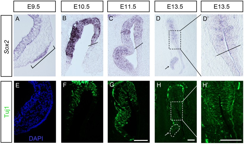
Fig. 1.
Sox2 and Tuj1 expression patterns in the olfactory epithelium of wild-type mouse. (A) At E9.5, expression of Sox2 is detected in the olfactory placode (bracket). (B-D′) From E10.5, Sox2 expression is restricted to the olfactory sensory epithelium. Black lines in B, C and D′ indicate the border between the sensory and respiratory epithelia. (E) At E9.5, no or only a few Tuj1+ neurons are detected in the olfactory placode. (F-H′) From E10.5, Tuj1+ neurons are located in the medial part of the sensory epithelium. (D,H) Sox2 and Tuj1 are also expressed in the vomeronasal organ, an olfactory epithelial derivative (indicated by arrows). D′ and H′ show higher magnifications of the boxed areas in D and H. Scale bars: 100 µm.

