Fig. 2.
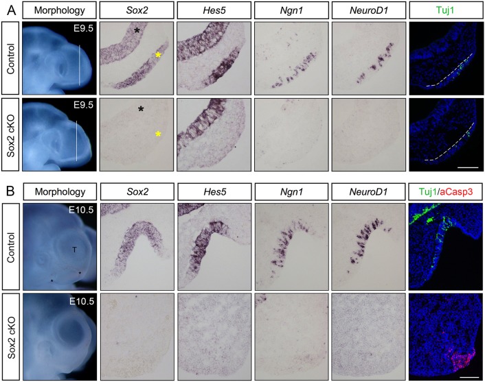
Loss of neurogenic markers in the Sox2-deficient olfactory epithelium. (A) At E9.5, cells in the control olfactory placode express Sox2. Hes5+ stem-like progenitors, Ngn1+ neuronal precursors, Neurod1+ and Tuj1+ post-mitotic neurons are detected in the olfactory placode (n=5). In E9.5 Sox2-deficient olfactory placodes, no expression of Sox2, Hes5, Ngn1 or Neurod1, and a reduced number of Tuj1+ cells are detected (n=4). The telencephalon is indicated by black asterisks, whereas the olfactory placode is marked by yellow asterisks, as well as by broken white lines. (B) At E10.5, the control olfactory epithelium is invaginated into a pit-like structure and sensory cells express Sox2; Hes5+ stem-like progenitors, Ngn1+ neuronal precursors, Neurod1+ and Tuj1+ post-mitotic neurons are detected in the epithelium (n=6). No or very few aCaspase3+ apoptotic cells are detected in the control olfactory epithelium (n=6). By E10.5, Sox2 cKO mutants (n=4) do not exhibit an olfactory epithelium, and no expression of any neurogenic markers is detected. A cluster of aCaspase3+ apoptotic cells is observed within the area of the disrupted olfactory epithelium. Scale bars: 100 µm.

