
Fig. 4.
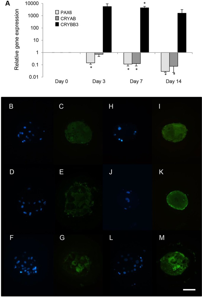
Aggregation of ROR1+ cells induces lens fibre cell crystallin expression. (A) Real-time PCR analysis of aggregated ROR1+ cells results in decreased relative expression of PAX6 and CRYAB, and increased expression of CRYBB3 (*P<0.01; data obtained from four biological replicates and presented as mean±s.e.m.). (B-M) Immunofluorescence analysis shows that after 14 days of culture, αA-crystallin (C) was expressed uniformly throughout the bulk of the micro-lenses, whereas β-crystallin (E) and γ-crystallin (G) were not. After 24 days of culture, αA-crystallin (I), β-crystallin (K) and γ-crystallin (M) were all expressed relatively uniformly throughout the bulk of the micro-lens. The location of DAPI-stained nuclei within the day 14 (B,D,F) and day 24 (H,J,L) aggregates are shown. Scale bar: 40 µm. Each image is representative of five micro-lenses from two biological replicates.