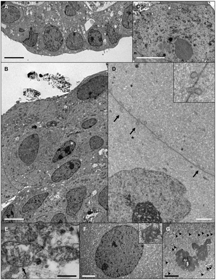
Fig. 5.
Evidence of progressive lens fibre cell differentiation in ROR1+ cell aggregates. Electron microscopy data from cultured aggregates. (A,B) A micro-lens cultured for 14 days shows a monolayer of LEC-like cells at the periphery of the tissue (A), and cells with small, rod-shaped nuclei (asterisk) and numerous organelles within the bulk of the tissue (B). (C) LEC-like cell with numerous organelles present at the periphery of a micro-lens after 24 days of culture. (D-G) Ultrastructural indicators of lens fibre cell differentiation within a micro-lens cultured for 42 days. (D) Ball-and-socket type membrane interdigitations (arrows) between adjacent lens fibre-like cells (inset shows a higher magnification of the region indicated with an arrow and asterisk). (E) A swollen mitochondria (arrow). (F) An enlarged nuclei with spoke-like nucleolus (inset). (G) A degraded nuclei with nuclear membrane visible as a series of vesicles (arrowheads). Scale bars: 5 µm in A-C; 2 µm in D,F,G; 0.5 µm in E. Images are representative of seven micro-lenses obtained from two biological replicates.

