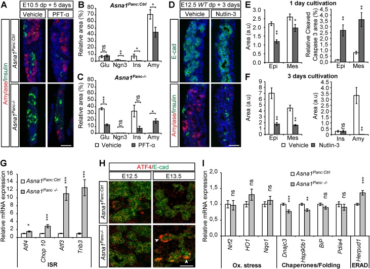
Fig. 3.
Apoptosis of pro-acinar progenitors in Asna1Panc−/− DPE is associated with p53 activity and activation of the IRS. (A) Representative immunohistochemistry of dorsal pancreas explants from E10.5 Asna1Panc:Ctrl and Asna1Panc−/− cultivated for 5 days exposed to vehicle (n=7 and 4, respectively) or PFT-α (30 µM) (n=11 and 4, respectively) using antibodies against amylase (red) and insulin (green). (B,C) Quantification of experiments described in A. Relative glucagon+ (Glu), Ngn3+, insulin+ (Ins) and amylase+ (Amy) areas over total E-cadherin+ area in explants from Asna1Panc:Ctrl (B) and Asna1Panc−/− (C) mice. (D) Representative immunohistochemistry of dorsal pancreas explants from E12.5 wild-type embryos cultivated for 3 days exposed to vehicle or Nutlin-3 (10 µM), using antibodies against E-cadherin (E-cad) and insulin (both green), and amylase (red). (E,F) Quantification of experiments described in D after 1 day (n=6 for each condition) (E) and 3 days (vehicle, n=4; Nutlin-3, n=5) (F) of cultivation; E-cadherin+ epithelial (Epi) and E-cadherin− mesenchymal (Mes) area; relative cleaved caspase 3+ (c.Casp.3) over E-cadherin+ area after 1 day; absolute insulin+ (Ins) and amylase+ (Amy) area after 3 days; arbitrary units (a.u). (G) qRT-PCR mRNA levels of the indicated ISR genes in dorsal pancreatic buds from E13.5 Asna1Panc:Ctrl (n=7) and Asna1Panc−/− (n=6) embryos. (H) Immunohistochemistry of dorsal pancreatic sections from E12.5 and E13.5 Asna1Panc:Ctrl and Asna1Panc−/− embryos (n=5) using antibodies against ATF4 (red) and E-cadherin (E-cad, green). Arrowheads indicate ATF4+ tip cells. (I) qRT-PCR mRNA levels of the indicated UPR genes in dorsal pancreatic buds from E13.5 Asna1Panc:Ctrl and Asna1Panc−/− (n=7) and Asna1Panc−/− (n=6) embryos. DAPI (blue) indicates nuclei in A,C. Scale bars: 50µm. Data are mean±s.e.m.; *P<0.05, **P<0.01, ***P<0.001; ns, not significant (Student's t-test).

