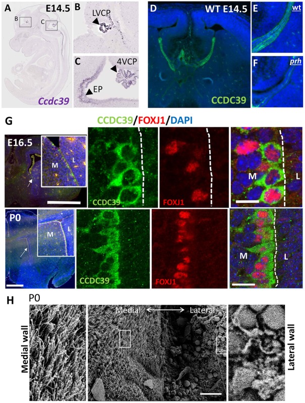
Fig. 4.
Ccdc39 expression in choroid plexus and ependymal epithelial cells of the prenatal mouse brain, and fully ciliated ependymal cells in the P0 mouse forebrain. (A-C) RNA in situ hybridization for Ccdc39 mRNA at E145 (adapted from Eurexpress.org). Ccdc39 is expressed (arrowheads) in the choroid plexus (CP) of the lateral ventricle (LV) and fourth ventricle (4V), as well as in ependymal cells (EP) and ependymal wall of the ventral fourth ventricle. (D-F) Immunohistochemistry with CCDC39 antibody of E14.5 wild-type (D,E) and prh mutant (F) brains. The CCDC39 protein is reduced to nearly undetectable levels in the prh mutant. (G) Immunohistochemistry with CCDC39 (green) and FOXJ1 (red) antibodies in E16 and P0 mouse brain. M, medial; L, lateral. Arrows indicate the location of the high-magnification images in the insets. (H) SEM images of ventromedial walls of P0 mouse forebrain showing fully ciliated ependymal cells. Magnifications of boxed areas are shown to each side. Scale bars: 1 mm in G (top left); 500 µm in G (bottom left); 5 µm in G (top right); 10 µm in G (bottom left); 50 µm in H.

