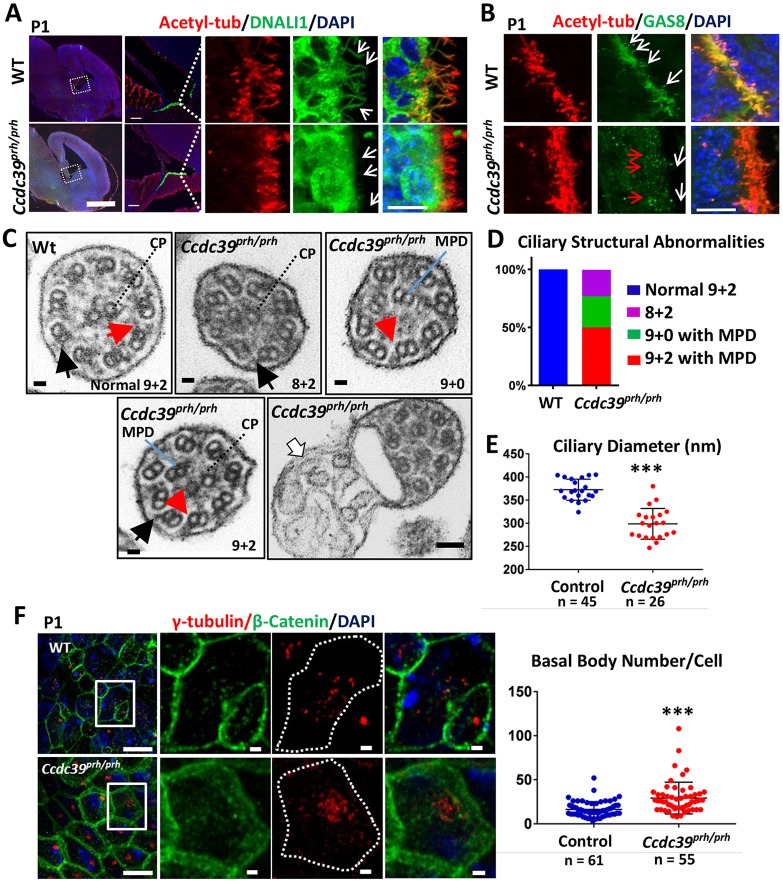
Fig. 6.
Ciliary structural abnormalities in ependymal cells of the Ccdc39prh/prh mutant mouse. (A) Immunofluorescence of the P1 brain for acetylated α-tubulin (red) and DNALI1 (green). Boxed regions are shown at higher magnification on the right. DNALI1 staining appears to colocalize with the acetylated α-tubulin staining within the axoneme of wild-type ependymal cells, but not in mutant (arrows). (B) Immunofluorescence of the P1 brain for acetylated α-tubulin (red) and GAS8 (green). An abnormal punctate staining pattern of GAS8 was detected in the apical side of the ependymal cells (red arrows), and no localization of GAS8 within the axoneme (white arrows) was found in the mutant. (C) TEM images of ependymal cilia in Ccdc39prh/prh and wild-type mice. A variety of microtubule disorganizations was found in the mutant, including absence of inner dynein arm (red arrows), mislocalization of one or two microtubule peripheral doublets (MPD), and absence of the central pair (CP). Outer dynein arms were preserved in the mutant (black arrows). Ectopic abnormal ciliary membrane inclusions were occasionally found in the mutant (white arrow). (D) About 27% (7/26) of mutant cilia lost the CP and showed MPD (green), ∼23% (5/26) lost MPD (purple), and ∼50% (13/26) showed normal CP and MPD (red). (E) Cross-sectional diameter of ependymal cilia was significantly reduced in the Ccdc39prh/prh mutant. ***P<0.0001. (F) P1 ependymal cilia stained for γ-tubulin (red) and β-catenin (green). Note the increased number of γ-tubulin-stained basal bodies in the Ccdc39prh/prh ependymal cells. Four sections from two animals each; ***P<0.00001. Scale bars: 1 mm in A (left); 100 µm in A (middle); 10 µm in A (right), B and F (right); 100 nm in C; 1 μm in F (left).

