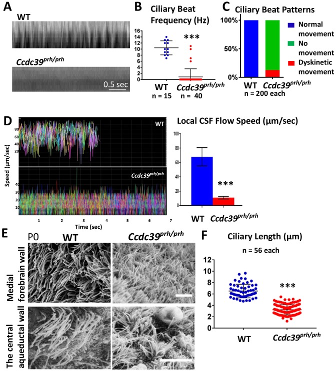
Fig. 7.
Abnormalities in ependymal cilia beating and CSF flow in Ccdc39prh/prh mice. (A) Representative kymograph of P7 wild-type and Ccdc39prh/prh mutant ependymal cilia from the high-speed video microscopy study. (B) Ciliary beat frequency was significantly reduced in the Ccdc39prh/prh mutant ependymal cilia. ***P<0.0001. (C) Ependymal ciliary beat patterns analyzed in the slowed down videos showed that mutant cilia were unable to generate a repetitive beating pattern (green) and only few (∼12%) showed dyskinetic movement (red). (D) The velocity of the fluorescent micro-beads analyzed from tracks in ex vivo P7 mouse brain ventricles of control and Ccdc39prh/prh mutants (see also Movies 7, 8). All micro-beads tested in the wild-type brain showed consistent speed at 68±0.7 µm/s and moved out of the field by 5 s of imaging. The speed of floating beads was significantly reduced in the Ccdc39prh/prh mice to 11±0.07 µm/s. More than 350 micro-beads were analyzed and are displayed in different colors in the graph. Two animals from each genotype were tested. Data represent mean±s.e.m. ***P<0.0001. (E) SEM images of multiciliated ependymal cells on the ventromedial forebrain wall and the central aqueduct wall of P0 mutant and wild-type mice. The Ccdc39prh/prh ependymal cells have shorter, thinner, but more abundant cilia. Scale bars: 5 µm. (F) Ependymal cilia length is significantly reduced in the Ccdc39prh/prh mutant. ***P<0.0001.

