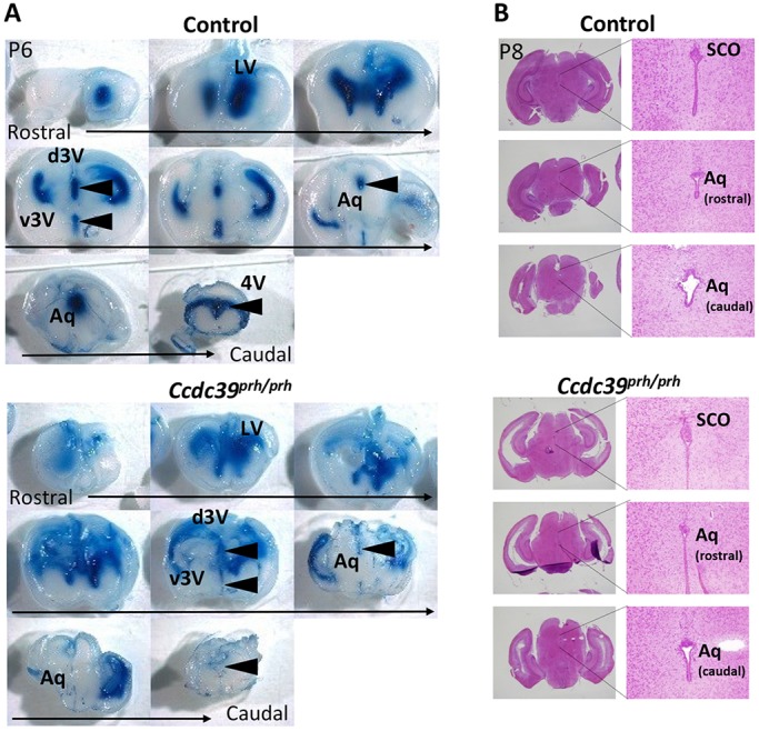
Fig. 8.

Retardation in CSF flow is not due to a physical obstruction in the central aqueduct of early postnatal prh mice. (A) CSF flow analysis in P6 Ccdc39prh/prh and control mice. Evans Blue dye injected into an anterior horn of the lateral ventricle (LV) traveled through the ventricular system and was detected in the third (3V) and fourth (4V) ventricles within 10 min in control mice (5/5) but not in the mutant (7/8) (arrowheads). (B) Histology of the central aqueduct (Aq) was comparable in mutant and littermate control mice at P8. SCO, subcommissural organ; d, dorsal; v, ventral.
