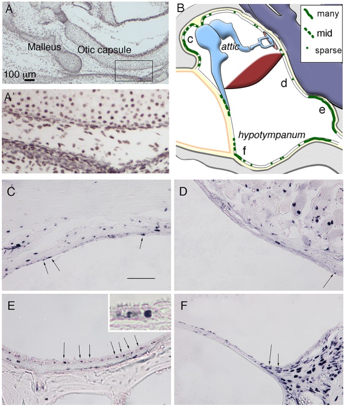
Fig. 2.

Label-retaining cells are concentrated in specific parts of the middle ear. (A,A′) Immunostained frontal section of a P1 mouse middle ear, following treatment of BrdU in the mother’s drinking water for 7 days using an antibody against BrdU. All cells are labelled in the ear. The boxed region in A is enlarged in A′. (B) Schematic highlighting distribution of BrdU-positive cells (green) within an example 8-week-old postnatal middle ear. Letters c-f represent regions shown in C-F. (C-F) Frontal sections of 8-week-old postnatal middle ear immunostained against BrdU, in the epithelium of the attic close to the ear drum (C), overlying the cochlea (D), in the hypotympanum close to the Eustachian tube (E) and near the ear drum on the ventral side (F). Arrows in C-F indicate positive epithelial cells. Inset in E shows labeling in basal cells. Scale bars: 100 µm in A; 100 µm in C-F.
