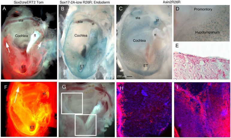
Fig. 3.
High levels of Sox2 expression and Wnt activity restricted to the endodermally derived part of the middle ear. (A-D) Whole-mount preparation of auditory bullae (AB) at P21 after ear drum removal showing the Eustachian tube (ET) ventrally (bottom) and stapes (st) dorsally (top). (E) Section of AB shown in C. (F-I) Whole-mount preparation of auditory bullae at 6 months. (A) Sox2creERTom mouse, tamoxifen injected 48 h prior to cull. Sox2-positive cells (and any progeny formed in the last 48 h) labelled in red. Arrows indicate tracts of red cells around the promontory. (B) Sox17-2A-icreR26R mouse, endoderm-derived cells and blood vessels stained blue. (C-E) Axin2R26R middle ears. Active Wnt signalling stains blue. (C) Whole bulla. (D) Positive scattered cells in hypotympanum near the ET, but not in the promontory overlying the cochlea. (E) Cryosection through the hypotympanum showing scattered blue cells in the epithelium, counterstained with Eosin. (F) AB; dark-field view showing Sox2-positive cells and progeny in orange. Tomato expression is also observed in the cochlea underlying the middle ear in stripes at 6 months. Arrow indicates Tomato-positive cells extending up to the attic around the promontory. (G) Dissected AB, boxed regions show areas highlighted by confocal microscopy in H,I. (H) Promontory and attic region with sparse isolated Tomato-positive cells. Large numbers of positive cells are observed at the margin of the promontory reaching up to the dorsal part of the middle ear. (I) Hypotympanum covered in Sox2-positive cells as at 2 days post-tamoxifen. The promontory (V-shape at top of image) only has a few positive cells in comparison. tt, tensor tympani muscle; st, stapes; ET, Eustachian tube; sta, stapedial artery running through stapes. Scale bar: 250 µm in A-C,F.

