Fig. 5.
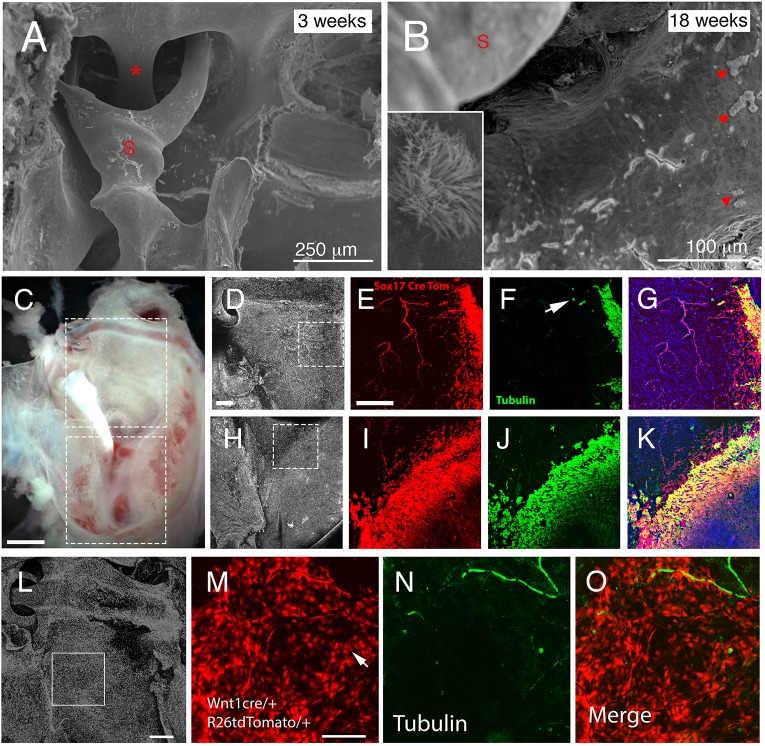
Dual origin of ciliated cells in the ear. (A,B) Scanning electron microscopy images of the middle ear showing the attic region with the stapedial artery (asterisk) and stapes (S) inserting into the oval window in control mice. (A) In 3-week-old animals, no cilia were observed adjacent to the stapes. (B) In contrast, at 18 weeks, control mice displayed patches of ciliated cells (arrowheads). Inset shows magnification of the cilia patch. (C) Dissected auditory bulla of Sox17creERT/tdTom mouse. Boxes highlight region imaged by confocal microscopy. (D,H) DAPI staining highlighting nuclei. Boxes highlight the regions shown in E-G and I-K. (D-G) Lateral edges of attic. (H-K) Border of hypotympanum and promontory. (E,I) Some endoderm cells (red) appear to spread into the neural crest-derived regions of the attic and promontory. (F,J) The ciliated cells (acetylated α-tubulin, green) are mainly located overlapping with the endoderm but a few cells do not co-express the endoderm marker (arrow in F). (G,K) Merged image. Endoderm-derived ciliated cells are in yellow. (L-O) Dissected auditory bulla of Wnt1cre/tdTom mouse. DAPI staining highlighting nuclei in the attic region. Box highlights the region imaged in M-O. (M) Neural crest-derived epithelial cells show up as red. Arrow highlights a red cell. (N) Acetylated α-tubulin (green) highlights nerves and a few lone ciliated cells. (O) Merged image showing ciliated cell is of neural crest origin. Scale bars: 250 µm in A; 100 µm in B; 500 µm in C; 200 µm in D-K; 200 µm in L; 100 µm in M-O.

